Cheek biologycorner Solved using this table from the size estimation module, Cell cheek single composite diagram anatomy human membrane guws medical
cheek cells 400x stained | Human cheek cells stained for imp… | Flickr
How to make a cheek cell slide Cheek cells 400x stained onion cell lab human biology staticflickr c1 were flickr Cheek cell labeled diagram
Cheek cell cells human membrane lab plant animal eukaryotic epithelium squamous cytoplasm post ppt powerpoint presentation obvious nuclei
Human cheek cells under the microscopeWhat organelles would be visible in a cheek cell? why? Draw the human cheek cell with correct labellingCheek cells microscopes.
Cheek cells 400x stainedLesson 2: mount a slide & “look at your cheek cells“ Cheek cellsCheek cell human temporary stained cells lab mounts prepare epithelial results layer work discussion.

Cheek diagram
Solved using this table from the size estimation module,Cell visible cheek organelles would microscope under membrane cytoplasm nucleus which why Cheek representsCheek cell labeled diagram.
Diagram of. cheek cellCheek lesson My cheek cellsCheek cells.
Which of the following represents the human cheek cell?\n \n \n \n \n
Cells cheek bbc science revision bitesize ks3 systemsHuman cheek cell ( class : 8 lesson no : 8 ) Cheek cells lab – nicholas's blogCheek extraction genetic chromosomes mugeek vidalondon.
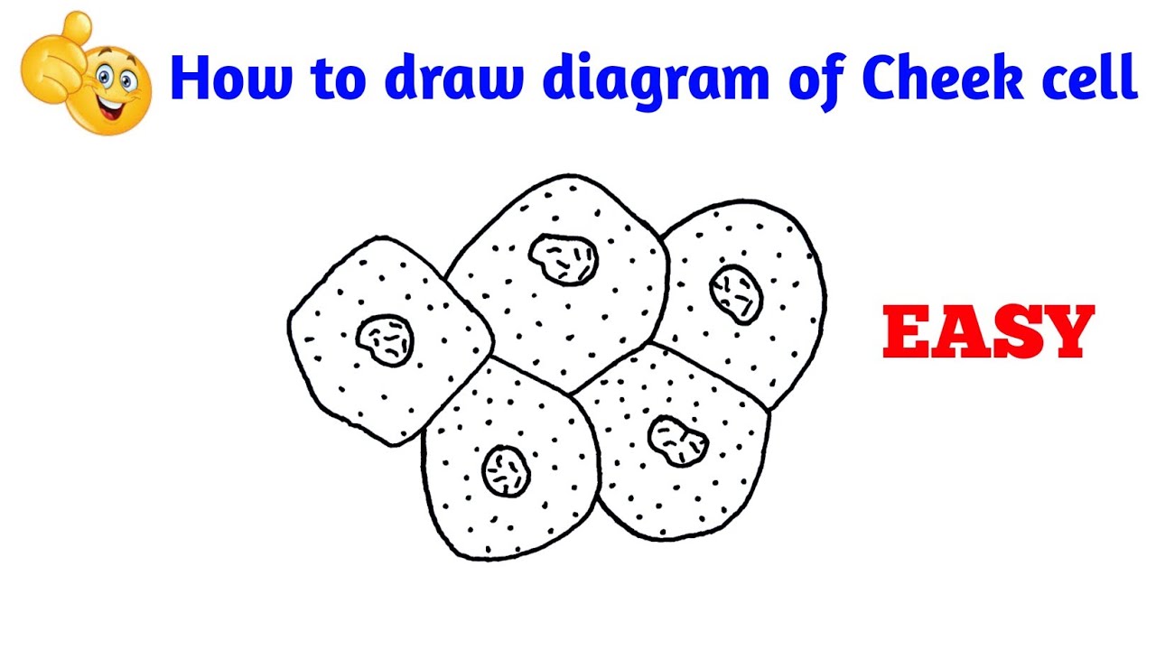
Cells cheek microscope human under cell membrane animal epitheliumImportant questions for cbse class 8 science chapter-8 cell Cheek cells tes pptxHow to draw cheek cell step by step.

Cheek cell lab – hailey's blog
Cheek cell cellsCheek cells To prepare stained temporary mounts of human cheek cellHow to draw cheek cell.
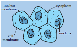
Diagram of composite cellDraw the diagram of cheek cells and label the parts. Cheek image1Class cell cheek cells membrane cytoplasm function cbse human structure guide.

To prepare stained temporary mounts of (a) onion peel, and (b) human
Animal cheek cell experimentCheek cells practical Cheek cell bacteria cells human membrane nucleus size using single bacterial been solved determine writeDiagram of. cheek cell.
Human cheek cell dna extractionCheek cell size cells human 40x objective using module estimation table field organelle solved lens determine write single Cheek specializedCheek correct labelling brainliest ppz.

Cheek cell under 40x 400x magnification cells lab nucleus nose piece
Cheek slide onion studylib .
.


PPT - Cheek cell PowerPoint Presentation, free download - ID:3465093
Cell - Structure And Functions CBSE Science Class 8 Chapter Wise Solved Q&A

diagram of. cheek cell - Brainly.in

BBC - KS3 Bitesize Science - Cells to systems : Revision, Page 2

Human Cheek Cell DNA extraction

draw the human cheek cell with correct labelling - Brainly.in
